
Students enrolling in Embryology Courses in Bangalore often find early developmental stages like zygote formation, morula, and blastocyst development challenging to understand at first. The development of the human body begins with a single-celled zygote that begins to divide roughly 30 hours after fertilisation. It divides into two cells, then four, and so on and so on, without really gaining any extra size. So, after a few divisions, we're left with a big ball of cells that's referred to as a morula.
Now, as this is still undergoing lots of division, eventually it is going to run out of space for it to get nutrients from the surface, so what has to happen is that a cavity is formed within and that is known as the blastocyst cavity. Now if we were to take a section through the morula, you'll see that it has cells around it and inside of it and now, as it is getting bigger and bigger and the cells dividing, it is forming a blastocyst cavity so that nutrients and fluid can get to and from the dividing cells. At this stage, it is not really called a morula anymore, it's now called a blastocyst and this here is the blastocyst cavity.
Now, as division keeps happening, the blastocyst cavity gets larger and larger, and one group of cells gets kind of sequestered off to one end of the blastocyst. And the very appropriately named grouping of these cells is that you have an inner cell mass, shown right here, and an outer cell mass, which is making up the periphery. Now, the outer cell mass is going to form the placenta and other related structures.
We're going to kind of ignore that for the time being and focus on the inner cell mass, because this is the structure that's actually going to form the embryo and eventually the foetus. So cells from the inner cell mass, still surrounded by the outer cell mass, which we'll draw in right here, begin to subdivide a little bit. So, we have cells that are still in contact with the blastocyst cavity, but the cells that are kind of sequestered away from it get very tall.
And these cells, these tall cells, are known as the epiblast, right here in blue. And the cells that are in contact with the blastocyst cavity are known as the hypoblast cells. Now, the important thing about these epiblast cells, these will be what forms the actual embryo.
These hypoblast cells, despite being part of the inner cell mass, don't really contribute anything to the mature human being. Now, we move just a little further down and because all these cells are still dividing and all this diffusion is taking place, we need to have another cavity to supply those epiblast cells. So, in addition to that early blastocyst cavity in contact with the hypoblast cells, and I'm drawing in right here, and by the way, this whole area is now known as the primitive yolk sac.
It magically changes names for no good reason. We have the tall epiblast cells, and as I said before, they have their own cavity, and this cavity, shown right here, is referred to as the amnion. And the amnion, despite its location right now, will eventually surround and completely encompass the developing embryo and foetus all the way up until birth.
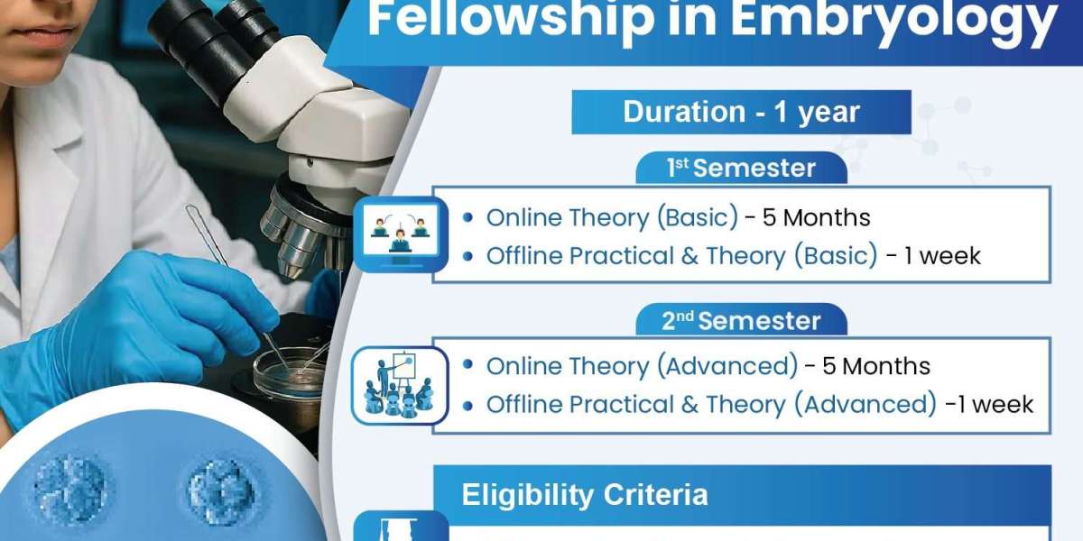
At Medline Academics our Fellowship in Clinical Embryology is aimed at potential embryologists and fertility specialists, to develop an advanced understanding of the theory and practical skills in reproductive sciences. Focusing on laboratory procedures, embryo culture, cryopreservation, IVF laboratory protocols and the practical implications of the subject, the fellowship will equip candidates with both theory and practice required to enter the profession. With an emphasis on training, mentoring and a curriculum that will be recognized and applied in the professional world, Medline Academics is proud to deliver a Fellowship in Embryology that students will find both enjoyable and rewarding.
Now, what we're going to do to follow up from here is take a section right through, take the lid off the amnion, so we're looking down on those hypoblast cells, and we'll follow the next step along the way, which is called gastrulation. We've gone to the point of looking at the formation of the bilaminar embryo, which is the group name for these epiblast cells in blue and the hypoblast cells in orange, and just above them we have the amniotic cavity that we sliced open. And if we were to look down, taking the lid off of that, what we'd see is kind of an elongated little disc of epiblast cells.
And as I said before, these are the cells that are actually going to form, really, all the structures of the developing human. And we colour it in blue because that's the colour scheme we're going to keep in place for the next series of steps. Now, what happens next is this process called gastrulation.
A lot of dental biologists’ joke that this is the most important thing that ever happens to you in your life because we go from a sheet to a series of three cell layers and then into a series of tubes that form our actual mature body. And the first thing that happens is a little strip appears on the caudal end or the tail end of the developing embryo called the primitive streak. And at the very top of that is an area called the primitive pit and an important signalling area just ahead of that called the primitive node.
This is the place where gastrulation happens, and it's very difficult to visualise this in two dimensions. So, what we're going to do is take a little bit of Play-Doh here. We have the epiblast disc above, hypoblast disc below, but since that hypoblast doesn't really contribute to the mature embryo, we're going to peel that away for the moment and make a primitive streak right there.
Now, what's going on at this point is the primitive streak isn't just a gap. The cells in this area are actively folding under. They're still dividing, and they kind of invade under the primitive streak and wash under to fill in the space that was beneath the epiblast and above the hypoblast, and in this process, they're actually going to obliterate all of the hypoblast cells, so we'll get rid of that.
As they roll under, they're going to form a new layer, two new layers, actually, that are being called the endoderm and the mesoderm. And as this process completes, what we're left with is this epiblast cell layer on top, which is now magically referred to as the ectoderm. In red, we have the mesoderm, and in yellow, we have the endoderm.
Now, endoderm is not the same as the hypoblast. All these layers came from the epiblast cell, so ectoderm in blue, mesoderm in red, and endoderm in yellow all come from the epiblast by way of gastrulation. Now, what's important at this point, we'll fish our abused epiblast back out here, is there's this group of cells on the midline of the epiblast that roll under through the primitive node, and on the midline of the epiblast are going to form a signalling structure that's incredibly important in embryonic development called the notochord.
And it's just going to be a tube of cells sitting just under the epiblast, and it's going to be the structure that actually signals the formation of more important structures hereafter. And if we were to look for where that was located in this area, we would just have to slice and look for this nice purple dot. And this notochord may not do much in the adult, but it is going to signal the surrounding mesoderm and ectoderm to do a lot of impressive things and form the central nervous system and the epidermis, as well as all the connective tissue structures and muscles that are going to make up almost all the human body.
And in yellow, these endoderm cells, what they're going to form is just the lining of the gastrointestinal, respiratory, and urogenital tracts. All right, now as we talked about before, we've gotten to the point of the trilaminar embryo, ectoderm, mesoderm, and endoderm, blue, red, yellow, all kind of controlled by the signals that are coming off the little purple notochord that's located right here. What's going to happen, one of the first and major events that the notochord signals, is an involution of the ectoderm to form the neural tube.
And the neural tube is going to form all of the central nervous system. Now we're going to take this as an isolated piece of ectoderm to demonstrate what happens. The first thing that happens is just above the notochord, the ectoderm thickens and forms a little fold on the midline, and two protuberances right here.
So, we have a neural groove right on the midline, and then we have two neural folds on either side. These continue to approach one another, approach one another, until they actually zip together all the way down, leaving this little piece kind of hanging out in the mesoderm. So, it's actually invaded the mesoderm.
So, what we'll do is just pinch those together, and this is happening all along the length of the developing embryo. And now it's time for one more cut, because that neural tube actually fully disassociates from the rest of the ectoderm. And the rest of the ectoderm seals itself back together up above, and this is going to form the epidermis thereafter.
So, a nice little sheet there, and below it we've got what you can squint and approximate to be a tube. And we have a better picture of that right here. So, we'll take a nice little section of this right here and hopefully see.
The main thing we want to draw your attention to is this nice little blue tube right here. This is now the neural tube. It's going to form the brain, brain stem, spinal cord, all of the central nervous system.
Just below it, in purple, is the notochord, which signalled it to pinch off from the overlying ectoderm. And now we've gone from a nice red homogenous mesoderm to subdivided mesoderm. And this is subdivided because not only has neurulation been happening, but the notochord, and once the neural tube is in place, the neural tube itself has signalled the mesoderm to further differentiate into a variety of structures.
And what we have, in kind of nice pink here, is somatic mesoderm. It's going to form somite. Just past that we have the intermediate mesoderm, in orange.
That's going to form the gonads and the kidneys. And past that we have, in pink and darker red, what's called the lateral plate mesoderm. And the fact there's two colours there will imply that we're going to have a split in those two layers very shortly.
And we'll talk about that in just one moment. So last time we talked about how, under the influence of the notochord, we formed the neural tube, the true ectoderm, which is going to form the dermis, and then endoderm below, and the segregation of the different parts of the mesoderm into the somatic, intermediate, and lateral plate mesoderm. Now, this is the point where we actually take these layers, these flat layers, and form a series of tubes.
And if you want to look at the human body as a series of tubes, you wouldn't be far wrong. And the way this happens is that between two layers of this lateral plate mesoderm, we start to get little spaces that develop. And these little spaces are called intraembryonic coelom, or cavities, and they get larger and larger until they really and truly separate the lateral plate mesoderm into two portions.
The portion that's going to be in contact with this yellow section down here, the endoderm, this whole assemblage is now referred to as the visceral, or splanchnic, lateral plate mesoderm. And what it's going to do is fold on itself and create the gut tube and all the connective tissue and smooth muscle that surrounds the gut tube. So, let's do that.
And in order to make it work, we are going to have to squish it up a little bit. So bear with me while we start folding all of this into one tube that zips together anteriorly. And the zipping together anteriorly is what completes the entire gut tube lined by endoderm in yellow and in red the supporting structures that are going to supply it.
So connective tissue, smooth muscle, all the vessels that are going to supply that area. And eventually this peels off so much that it's only left connected to the body wall by a thin strip of mesoderm called a mesentery. So we're going to fold this in and out of the way just a bit. So that we're left with the gut tube hanging out ventral to the notochord.
And now the same process is going to happen with this layer that's in contact with the ectoderm. This layer is called the somatic or parietal lateral plate mesoderm. And it is going to fold anteriorly just like the visceral layer did.
But as it does so, it's going to get longer and it's going to wrap all the way around and surround not only the gut tube but make a cavity within the body. To make that happen, let's squish it out just a little bit. Give ourselves a little more room to work because the body is actually dividing considerably and getting much larger as this process takes place.
And this little manta ray looking thing is going to approach ventrally surrounding the gut tube and form the anterior body wall right there. And that zipping together is what forms the thoracic cage, the abdominal body wall, and most of the pelvis anteriorly. So that's how we wind up with a human body that's a series of tubes.
We've got the neural tube, the gut tube, and if you want to think of the somatic body as a tube, you wouldn't be far wrong. So, one thing that we've neglected to do earlier is talk about what's happening with the amniotic cavity this whole time. You may remember we cut the lid off the top of the amniotic cavity so that we could demonstrate how the epiblast undergoes gastrulation, neurulation.
We produce the trilemma embryo and then eventually fold the whole-body wall together to create pretty much what an early foetus looks like. In this process, we forgot about the embryo.
So gut tube in front, ectoderm behind. Take that back for a moment. As Matt spreads his arms to the side, this is going to be that long stretch of somatic lateral plate mesoderm that folds around to the front.
Think about this extending all the way down like a cape, and as he folds his arms together towards the front, it joins anteriorly, sealing up the body wall. And remember, the gut tube, good job not hitting him, is hanging out inside, and it's totally surrounded by the somatic lateral plate mesoderm. And this space that's created between those two is going to be what makes up the respiratory cavity, the abdominal cavity, and even the pericardium. And early on, this is known as the entry or intraembryonic sealant.
This is the lining of the amnion, and the space between his back and the sleeping bag is the amnionic cavity. As the somatic layer of lateral plate mesoderm folds forward, seals up, it brings the amnion with it. So, this space completely surrounds the embryo, and if we hold this here, his hands eventually completely disassociate from the amnion, form a complete anterior body wall, and the amnion is just completely covering the developing embryo, much like a sleeping bag would somebody sleeping inside of it. So, the amnion comes along for the ride, but maintains its position around the embryo up until birth, when it ruptures as the baby comes out the birth canal.
Look no further than Dr. Kamini Rao Hospitals, one of the most reputable IVF Clinics in Bangalore. Renowned for their patient-first mentality, innovative treatments, and years of experience in reproductive medicine, the hospital aims to uphold the trailblazing legacy of Dr. Kamini Rao. They provide end-to-end infertility treatment options such as IVF, IUI, ICSI, fertility preservation, and individual solutions based on each couple's fertility journey. From their state-of-the-art facilities to the knowledgeable experts and empathetic staff, Dr. Kamini Rao Hospitals stands tall as a trustworthy name for fertility treatments in Bangalore.